Cellular injury ( part 2) apoptosis and necrosis l General pathology revision for dental students
to see first part click here
to download full pdf click here
Cellular damage and disappearance :
- May be necrosis (Groups of cells or tissue) or apoptosis (Single cell or sometimes few cells).
NECROSIS
- Local death of cells or tissues within the living body. Necrosis occurs either directly or follows reversible injury.
Causes :
- hypoxia, toxins , viral hepatitis, HIV, hypersensitivity , free radical as O2
Pathogenesis :
- Severe
mitochondrial damage →marked
↓of
ATP production with metabolic shift to anaerobic glycolysis →↓intracellular PH
(acidosis → loss
of integrity of the cellular membranes as disruption of lysosomal membranes and
lysosomal enzymes set free.)
- Increase
intracellular Ca: ↓ATP
→failure
of Ca pump (Ca is normally maintained at extremely low levels) Increased Ca
leads to activation of the following enzymes: (a. Proteases leading to
breakdown of cytoskeleton protein), (Phospholipases leading to destruction of
membrane phospholipids), (Endonucleases leading to breakdown of DNA and
chromatin fragmentation
- these lead to protein denaturation &release enzymatic digestion(cellular or polymorph lysosome)
Macroscopic Picture :
- Necrotic tissue appears opaque and whitish or yellowish in color. The surrounding tissue appears red due to inflammatory hyperemia.
Microscopic Picture :
- Immediately after necrosis dead cells appear like living cells. Then a series of changes
Nuclear Changes:
- Pyknosis : The nucleus shrinks , its chromatin becomes dense and it stains darkly.
- Karyorrhexis: The nucleus breaks up into multiple small fragments. Commonly a pyknotic nucleus undergoes karyorrhexis.
- Karyolysis: The nucleus appears to dissolve and fails to take the stain due to chromatin hydrolysis.
Cytoplasmic Changes:
- The cells appear swollen (cytomegaly).
- In haematoxylin and eosin stained sections there is cytoplasmic eosinophilia. This is due to loss of RNA with consequent loss of the normal cytoplasmic basophilia, plus increased binding of eosin to denatured cytoplasmic proteins.
- The cells loose the cell membrane and become indistinct from each other.
- Blue stained calcium deposit appear later on.
Types of Necrosis:
Coagulative Necrosis:
- Commonly caused by sudden cut of the blood supply as in infarcts. It seems likely that the cellular proteins are denatured causing the tissue to become firm and opaque white.
- Microscopically: Early the general architecture of the tissue is seen but with no cellular details. Lately dead tissue appears homogenous, structureless and pink stained. The blood vessels and fibrous stroma are more resistant to the process of necrosis and their outlines remain for a longer time.
Liquefactive Necrosis:
- The necrotic tissue is rapidly liquefied . It occurs in:
- Infarctions of the brain and spinal cord: The softening and liquefaction is due to the high lipid and fluid content of the nervous tissue.
- Pyogenic abscess: The central necrotic core is liquefied by the proteolytic enzymes released from the pus cells.
- Amoebic abscess: The liquefaction is due to the action of liquefactive enzymes produced by the parasite.
Caseation Necrosis:
- Necrosis followed by slow partial liquefaction.
- Grossly : the caseating material is dry, pale yellow and resembles cream cheese or casein.
- Microscopically all cellular details are lost and the tissues appear structureless , granular or homogenous and pink stained.
- Caseation occurs in tuberculous lesions and in gumma of syphilis.
- Caseation in gumma is slow and incomplete, so the outline of the tissue is preserved for a longer time. Caseation necrosis is caused by an antigen-antibody reaction(allergic necrosis).
Fat Necrosis: Is of two types:
Enzymatic fat necrosis: It
occurs in acute hemorrhagic pancreatitis.
- The enzyme lipase which escapes from the ruptured pancreatic ducts acts on the fat of the omentum, mesentery and abdominal organs.
- Lipase splits fat into glycerol and fatty acids. Glycerol is absorbed in the blood. Fatty acids deposit with calcium as small dull opaque white patches. Microscopically the affected fat cells appear cloudy and surrounded by foreign body giant cell reaction and fibrosis.
Traumatic fat necrosis:
- It occurs as a result of trauma to the adipose tissue of the breast and subcutaneous fat . The fat cells rupture and self-digestion takes place.
Fate of Necrotic Tissue:
- Small area of necrosis: Part
of the necrotic tissue is removed by the macrophages. The rest gets liquefied
and drained by the lymphatics and veins. Healing occurs by regeneration or by granulation
tissue formation followed by fibrosis.
- Large area of necrosis: Gets
surrounded by a fibrous capsule and may show dystrophic calcification later on.
APOPTOSIS
- Distinctive morphologic pattern of cell death affecting a single cell or small group of cells. Apoptosis is an energy-dependent programmed
- cell death for removal of unwanted individual cells. Apoptosis literally in Greek means "dropping off".
Morphological Changes:
- Shrinkage of the cell.
- Condensation and fragmentation of chromatin.
- Rapid break down of the cell to form apoptotic bodies. Apoptotic bodies have intense eosinophilic cytoplasm and dense chromatin fragments. Apoptotic bodies are phagocytosed by neighboring cells or by macrophages.
- Lack of inflammation in surrounding tissues.
Occurrence:
- Occur in normal cell turnover.
- Programmed cell destruction during embryonic development (e.g.in the interdigital clefts).
- Endocrine dependent involution of tissues e.g. shedding of the endometrium during the menstrual cycle and regression of lactating breast after weaning.
- In pathological conditions e.g.:
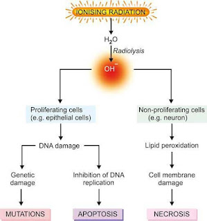
- Radiation cell injury.
- Cell death by cytotoxic T lymphocytes.
- Liver cells in viral hepatitis.
- Reduction of cell number in pathological atrophy.


0 Comments